How the Human Mind Really Works
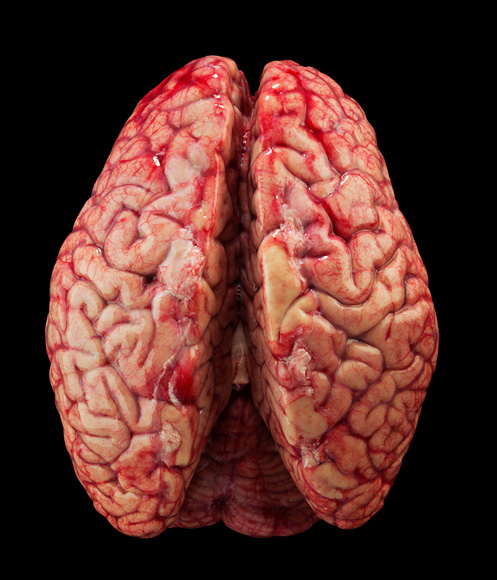
Photograph by Robert Clark; brain preparation performed at Allen Institute for Brain Science
Centuries of study have provided increasingly detailed understanding of human brain anatomy.
Photograph by Robert Clark
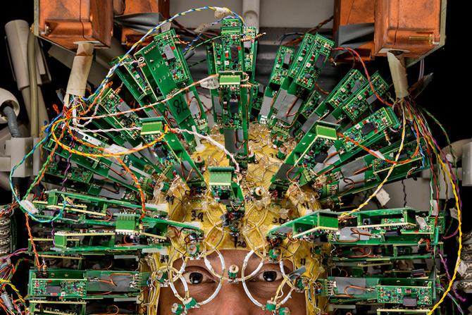
Mind Machine
An engineer wears a helmet of sensors at the Martinos Center for Biomedical Imaging—part of a –
brain scanner requiring almost as much power as a nuclear submarine.
Antennas pick up signals produced when the scanner’s magnetic field excites water molecules in the brain.
Computers convert this data into brain maps like the one in the following image.
Photograph by Robert Clark; brain preparation performed at Allen Institute for Brain Science. Image by Van Weeden and L. L. Wald, Martinos Center for Biomedical Imaging, Human Connectome Project
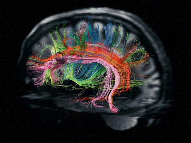
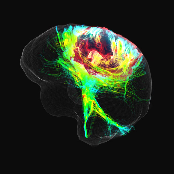
The Color of Thought
Scientists are turning their attention to the complex circuits that connect the brain’s many
regions—some 100,000 miles of fibers called white matter, enough to circle the Earth four times.
In this image taken at the Martinos Center, pink and orange bundles transmit signals critical for language.
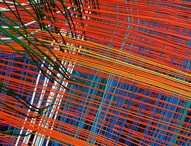
Image by Van Weeden and L. L. Wald, Martinos Center for Biomedical Imaging, Human Connectome Project
Anatomy of a Mystery
New technologies let scientists peer deep into the hidden structure of the brain.
A high-resolution view of the image above reveals white matter fibers arranged in a
mysterious grid structure, like longitude and latitude lines on a map.
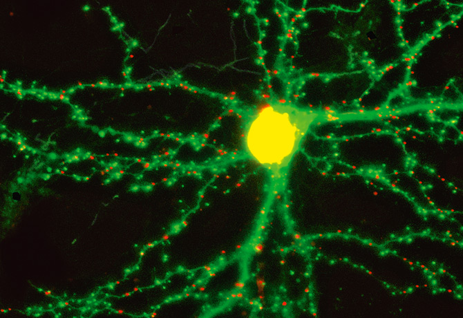
Image by Garrett Gross and Don Arnold, University of Southern California
The Glow of Memory
When you form a memory, “there’s a physical change in the brain,”
says Don Arnold, of the University of Southern California.
Red and green dots on the branches extending from this rat neuron show where it contacts other neurons.
As the rat forms new memories, new dots appear and old ones vanish.
Photograph by Robert Clark
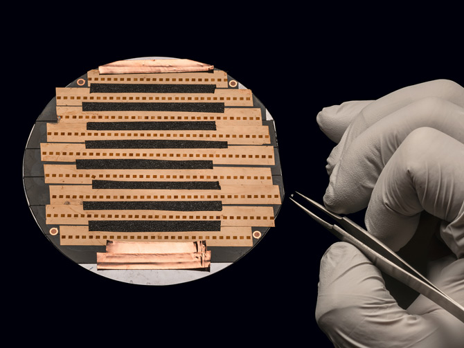
Intimate View
Two hundred sections of a piece of mouse brain, each less than 1/1,000
the thickness of a human hair, are readied to be imaged by an electron microscope.
Arranged in stacks, 10,000 such photomicrographs form a 3-D model no larger than a grain of salt (in tweezers).
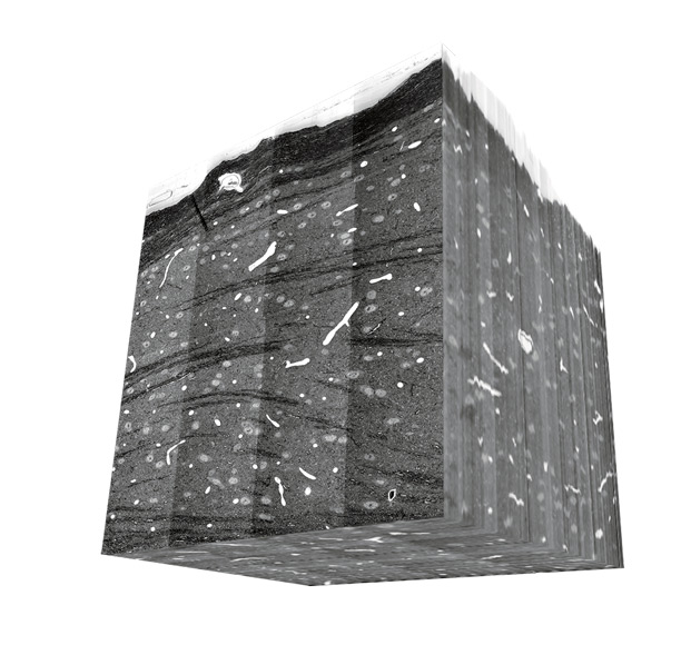
Image by Josh L. Morgan, Harvard University; Arthur Wetzel, Pittsburgh Supercomputing Center
Intimate View
Arranged in stacks, 10,000 photomicrographs form a 3-D model no larger than a grain of salt.
A human brain visualized at this level of detail would require an amount of data
equal to all the written material in all the libraries of the world.

Jennifer on the Brain
Caltech and UCLA scientists use pictures of celebrities to study how the brain processes what the eyes see.
In 2005 they found an individual nerve cell that fired only when subjects were shown pictures of Jennifer Aniston.
Another neuron responded only to pictures of Halle Berry—even when she was masked as Catwoman.
Follow-up studies suggest that relatively few neurons are involved in representing any given person, place,
or concept, making the brain staggeringly efficient at storing information.
Photograph by Rob Clark
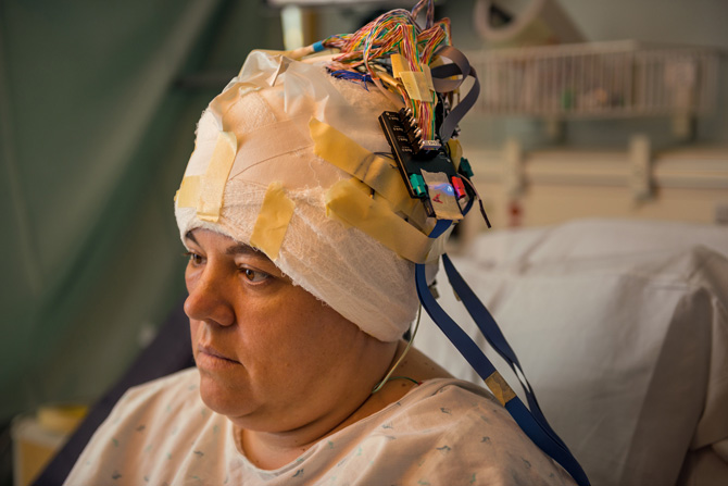
Listening In
How did scientists discover the “Jennifer Aniston neuron”? At UCLA’s Medical Center for Neuroscience
electrodes are implanted in the brains of epileptic patients such as Crystal Hawkins.
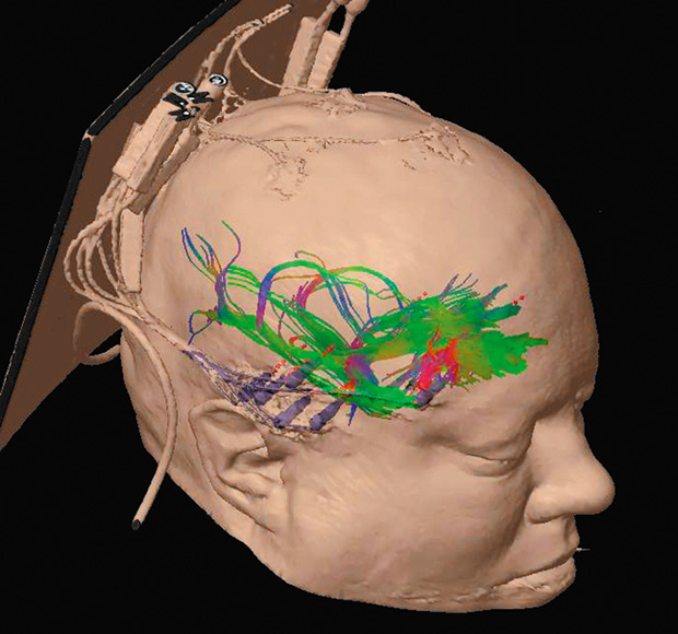
Image by Eric Behnke and Andrew Frew, UCLA
Listening In
The next time Hawkins has a seizure, the electrodes (purple cones) will pinpoint its source,
allowing neurosurgeons to target what brain tissue to remove.
The electrodes also provide a rare opportunity to eavesdrop on neurons functioning normally,
which led to the discovery of nerve cells that respond to specific faces.
Photograph by Robert Clark
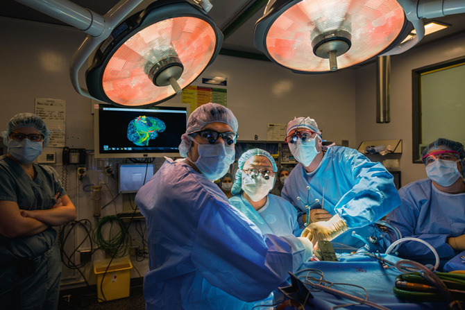
No Room for Error
Removing brain tumors is a risky procedure—surgeons need to excise as much of a tumor as possible without destroying
neurons essential for functions such as speech, sight, and memory or the connective fibers between them.
David Fortin (at center right), a neurosurgeon at the Université de Sherbrooke in Canada,
relies on a high-resolution map of a patient’s brain to avoid mishaps.
Maxime Descoteaux and Maxime Chamberland, Sherbrooke Connectivity Imaging Lab, Université de Sherbrooke
A Guided Hand
Scans of one of Fortin’s patients revealed that a tumor (red) had grown
into a region controlling the movement of hands and feet.
Photograph by Robert Clark
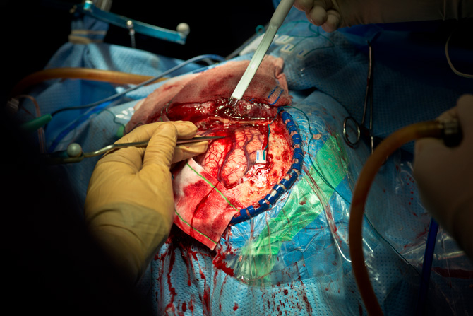
Critical Cuts
As he removed parts of the tumor, Fortin applied current to the region to determine if
neighboring neurons were critical for movement. “There was a lot of motor function still active in this patient,”
says Maxime Descoteaux, one of the Université de Sherbrooke scientists who made the brain scan.
“So the surgeon in this case was more conservative than aggressive.”
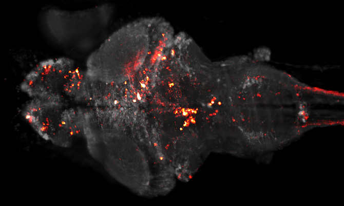
Image by Philipp Keller and Misha Ahrens, Janelia Farm (HHMI)
Lighting the Way
Scientists at the Janelia Farm Research Campus of the Howard Hughes Medical Institute
have added a gene to the neurons in zebrafish brains that causes them to produce light each time they send a signal.
Because zebrafish are transparent, the scientists can watch the sparkling activity of most of the 100,000 neurons in the animal’s brain.
The patterns of flashes are giving them new insights into how all brains process information.
Photo Credits: http://www.robertclarkphoto.com/
Source Site: http://ngm.nationalgeographic.com/2014/02/brain/clark-photography