What Is the Heart?
Your heart is a muscular organ that pumps blood to your body. Your heart is at the center of your circulatory system. This system consists of a network of blood vessels, such as arteries, veins, and capillaries. These blood vessels carry blood to and from all areas of your body.
An electrical system controls your heart and uses electrical signals to contract the heart’s walls. When the walls contract, blood is pumped into your circulatory system. Inlet and outlet valves in your heart chambers ensure that blood flows in the right direction.
Your heart is vital to your health and nearly everything that goes on in your body. Without the heart’s pumping action, blood can’t move throughout your body.
Your blood carries the oxygen and nutrients that your organs need to work well. Blood also carries carbon dioxide (a waste product) to your lungs so you can breathe it out.
A healthy heart supplies your body with the right amount of blood at the rate needed to work well. If disease or injury weakens your heart, your body’s organs won’t receive enough blood to work normally.
Your heart is located under your ribcage in the center of your chest between your right and left lungs. Its muscular walls beat, or contract, pumping blood to all parts of your body.
The size of your heart can vary depending on your age, size, and the condition of your heart. A normal, healthy, adult heart usually is the size of an average clenched adult fist. Some diseases can cause the heart to enlarge.
The Exterior of the Heart
Below is a picture of the outside of a normal, healthy, human heart.
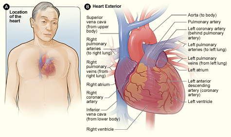
Figure A shows the location of the heart in the body. Figure B shows the front surface of the heart, including the coronary arteries and major blood vessels.
In figure B, the heart is the muscle in the lower half of the picture. The heart has four chambers. The heart’s upper chambers, the right and left atria (AY-tree-uh), are shown in purple. The heart’s lower chambers, the right and left ventricles (VEN-trih-kuls), are shown in red.
Some of the main blood vessels (arteries and veins) that make up your circulatory system are directly connected to the heart.
The Right Side of Your Heart
In figure B above, the superior and inferior vena cavae are shown in blue to the left of the heart muscle as you look at the picture. These veins are the largest veins in your body.
After your body’s organs and tissues have used the oxygen in your blood, the vena cavae carry the oxygen-poor blood back to the right atrium of your heart.
The superior vena cava carries oxygen-poor blood from the upper parts of your body, including your head, chest, arms, and neck. The inferior vena cava carries oxygen-poor blood from the lower parts of your body.
The oxygen-poor blood from the vena cavae flows into your heart’s right atrium and then to the right ventricle. From the right ventricle, the blood is pumped through the pulmonary (PULL-mun-ary) arteries (shown in blue in the center of figure B) to your lungs.
Once in the lungs, the blood travels through many small, thin blood vessels called capillaries. There, the blood picks up more oxygen and transfers carbon dioxide to the lungs—a process called gas exchange. To learn more about gas exchange, go to the Health Topics How the Lungs Work article.
The oxygen-rich blood passes from your lungs back to your heart through the pulmonary veins (shown in red to the left of the right atrium in figure B).
The Left Side of Your Heart
Oxygen-rich blood from your lungs passes through the pulmonary veins (shown in red to the right of the left atrium in figure B above). The blood enters the left atrium and is pumped into the left ventricle.
From the left ventricle, the oxygen-rich blood is pumped to the rest of your body through the aorta. The aorta is the main artery that carries oxygen-rich blood to your body.
Like all of your organs, your heart needs oxygen-rich blood. As blood is pumped out of your heart’s left ventricle, some of it flows into the coronary arteries (shown in red in figure B).
Your coronary arteries are located on your heart’s surface at the beginning of the aorta. They carry oxygen-rich blood to all parts of your heart.
The Interior of the Heart
Below is a picture of the inside of a normal, healthy, human heart.
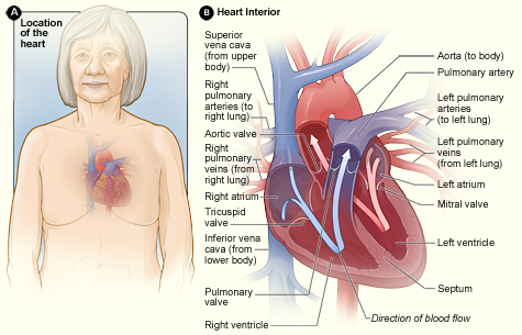
Figure A shows the location of the heart in the body. Figure B shows a cross-section of a healthy heart and its inside structures. The blue arrow shows the direction in which oxygen-poor blood flows through the heart to the lungs. The red arrow shows the direction in which oxygen-rich blood flows from the lungs into the heart and then out to the body.
Heart Chambers
Figure B shows the inside of your heart and how it’s divided into four chambers. The two upper chambers of your heart are called the atria. They receive and collect blood.
The two lower chambers of your heart are called ventricles. The ventricles pump blood out of your heart to other parts of your body.
The Septum
An internal wall of tissue divides the right and left sides of your heart. This wall is called the septum.
The area of the septum that divides the atria is called the atrial or interatrial septum. The area of the septum that divides the ventricles is called the ventricular or interventricular septum.
Heart Valves
Figure B shows your heart’s four valves. Shown counterclockwise in the picture, the valves include the aortic (ay-OR-tik) valve, the tricuspid (tri-CUSS-pid) valve, the pulmonary valve, and the mitral (MI-trul) valve.
Blood Flow
The arrows in figure B show the direction that blood flows through your heart. The light blue arrow shows that blood enters the right atrium of your heart from the superior and inferior vena cavae.
From the right atrium, blood is pumped into the right ventricle. From the right ventricle, blood is pumped to your lungs through the pulmonary arteries.
The light red arrow shows oxygen-rich blood coming from your lungs through the pulmonary veins into your heart’s left atrium. From the left atrium, the blood is pumped into the left ventricle. The left ventricle pumps the blood to the rest of your body through the aorta.
For the heart to work well, your blood must flow in only one direction. Your heart’s valves make this possible. Both of your heart’s ventricles have an “in” (inlet) valve from the atria and an “out” (outlet) valve leading to your arteries.
Healthy valves open and close in exact coordination with the pumping action of your heart’s atria and ventricles. Each valve has a set of flaps called leaflets or cusps that seal or open the valve. This allows blood to pass through the chambers and into your arteries without backing up or flowing backward.
Heart Contraction and Blood Flow
The animation below shows how your heart pumps blood. Click the “start” button to play the animation. Written and spoken explanations are provided with each frame. Use the buttons in the lower right corner to pause, restart, or replay the animation, or use the scroll bar below the buttons to move through the frames.
Heartbeat
Almost everyone has heard the real or recorded sound of a heartbeat. When your heart beats, it makes a “lub-DUB” sound. Between the time you hear “lub” and “DUB,” blood is pumped through your heart and circulatory system.
A heartbeat may seem like a simple, repeated event. However, it’s a complex series of very precise and coordinated events. These events take place inside and around your heart.
Each side of your heart uses an inlet valve to help move blood between the atrium and ventricle. The tricuspid valve does this between the right atrium and ventricle. The mitral valve does this between the left atrium and ventricle. The “lub” is the sound of the tricuspid and mitral valves closing.
Each of your heart’s ventricles also has an outlet valve. The right ventricle uses the pulmonary valve to help move blood into the pulmonary arteries. The left ventricle uses the aortic valve to do the same for the aorta. The “DUB” is the sound of the aortic and pulmonary valves closing.
Each heartbeat has two basic parts: diastole (di-AS-toe-lee) and systole (SIS-toe-lee).
During diastole, the atria and ventricles of your heart relax and begin to fill with blood. At the end of diastole, your heart’s atria contract (atrial systole) and pump blood into the ventricles.
The atria then begin to relax. Next, your heart’s ventricles contract (ventricular systole) and pump blood out of your heart.
Pumping Action
Your heart uses its four valves to ensure your blood flows in only one direction. Healthy valves open and close in coordination with the pumping action of your heart’s atria and ventricles.
Each valve has a set of flaps called leaflets or cusps that seal or open the valve. The cusps allow pumped blood to pass through the chambers and into your blood vessels without backing up or flowing backward.
Oxygen-poor blood from the vena cavae fills your heart’s right atrium. The atrium contracts (atrial systole). The tricuspid valve located between the right atrium and ventricle opens for a short time and then shuts. This allows blood to enter the right ventricle without flowing back into the right atrium.
When your heart’s right ventricle fills with blood, it contracts (ventricular systole). The pulmonary valve located between your right ventricle and pulmonary artery opens and closes quickly.
This allows blood to enter into your pulmonary arteries without flowing back into the right ventricle. This is important because the right ventricle begins to refill with more blood through the tricuspid valve. Blood travels through the pulmonary arteries to your lungs to pick up oxygen.
Oxygen-rich blood returns from the lungs to your heart’s left atrium through the pulmonary veins. As your heart’s left atrium fills with blood, it contracts. This event is called atrial systole.
The mitral valve located between the left atrium and left ventricle opens and closes quickly. This allows blood to pass from the left atrium into the left ventricle without flowing backward.
As the left ventricle fills with blood, it contracts. This event is called ventricular systole. The aortic valve located between the left ventricle and aorta opens and closes quickly. This allows blood to flow into the aorta. The aorta is the main artery that carries blood from your heart to the rest of your body.
The aortic valve closes quickly to prevent blood from flowing back into the left ventricle, which already is filling up with new blood.
Taking Your Pulse
When your heart pumps blood through your arteries, it creates a pulse that you can feel on the arteries close to the skin’s surface. For example, you can feel the pulse on the artery inside of your wrist, below your thumb.
You can count how many times your heart beats by taking your pulse. You will need a watch with a second hand.
To find your pulse, gently place your index and middle fingers on the artery located on the inner wrist of either arm, below your thumb. You should feel a pulsing or tapping against your fingers.
Watch the second hand and count the number of pulses you feel in 30 seconds. Double that number to find out your heart rate or pulse for 1 minute.
The usual resting pulse for an adult is 60 to 100 beats per minute. To find your resting pulse, count your pulse after you have been sitting or resting quietly for at least 10 minutes.
Your heart and blood vessels make up your overall blood circulatory system. Your blood circulatory system is made up of four subsystems.
Arterial Circulation
Arterial circulation is the part of your circulatory system that involves arteries, like the aorta and pulmonary arteries. Arteries are blood vessels that carry blood away from your heart. (The exception is the coronary arteries, which supply your heart muscle with oxygen-rich blood.)
Healthy arteries are strong and elastic (stretchy). They become narrow between heartbeats, and they help keep your blood pressure consistent. This helps blood move through your body.
Arteries branch into smaller blood vessels called arterioles (ar-TEER-e-ols). Arteries and arterioles have strong, flexible walls that allow them to adjust the amount and rate of blood flowing to parts of your body.
Venous Circulation
Venous circulation is the part of your circulatory system that involves veins, like the vena cavae and pulmonary veins. Veins are blood vessels that carry blood to your heart.
Veins have thinner walls than arteries. Veins can widen as the amount of blood passing through them increases.
Capillary Circulation
Capillary circulation is the part of your circulatory system where oxygen, nutrients, and waste pass between your blood and parts of your body.
Capillaries are very small blood vessels. They connect the arterial and venous circulatory subsystems.
The importance of capillaries lies in their very thin walls. Oxygen and nutrients in your blood can pass through the walls of the capillaries to the parts of your body that need them to work normally.
Capillaries’ thin walls also allow waste products like carbon dioxide to pass from your body’s organs and tissues into the blood, where it’s taken away to your lungs.
Pulmonary Circulation
Pulmonary circulation is the movement of blood from the heart to the lungs and back to the heart again. Pulmonary circulation includes both arterial and venous circulation.
Oxygen-poor blood is pumped to the lungs from the heart (arterial circulation). Oxygen-rich blood moves from the lungs to the heart through the pulmonary veins (venous circulation).
Pulmonary circulation also includes capillary circulation. Oxygen you breathe in from the air passes through your lungs into your blood through the many capillaries in the lungs. Oxygen-rich blood moves through your pulmonary veins to the left side of your heart and out of the aorta to the rest of your body.
Capillaries in the lungs also remove carbon dioxide from your blood so that your lungs can breathe the carbon dioxide out into the air.
Your Heart’s Electrical System
The animation below shows how your heart’s electrical system works. Click the “start” button to play the animation. Written and spoken explanations are provided with each frame. Use the buttons in the lower right corner to pause, restart, or replay the animation, or use the scroll bar below the buttons to move through the frames.
Your heart’s electrical system controls all the events that occur when your heart pumps blood. The electrical system also is called the cardiac conduction system. If you’ve ever seen the heart test called an EKG (electrocardiogram), you’ve seen a graphical picture of the heart’s electrical activity.
Your heart’s electrical system is made up of three main parts:
- The sinoatrial (SA) node, located in the right atrium of your heart
- The atrioventricular (AV) node, located on the interatrial septum close to the tricuspid valve
- The His-Purkinje system, located along the walls of your heart’s ventricles
A heartbeat is a complex series of events. These events take place inside and around your heart. A heartbeat is a single cycle in which your heart’s chambers relax and contract to pump blood. This cycle includes the opening and closing of the inlet and outlet valves of the right and left ventricles of your heart.
Each heartbeat has two basic parts: diastole and systole. During diastole, the atria and ventricles of your heart relax and begin to fill with blood.
At the end of diastole, your heart’s atria contract (atrial systole) and pump blood into the ventricles. The atria then begin to relax. Your heart’s ventricles then contract (ventricular systole), pumping blood out of your heart.
Each beat of your heart is set in motion by an electrical signal from within your heart muscle. In a normal, healthy heart, each beat begins with a signal from the SA node. This is why the SA node sometimes is called your heart’s natural pacemaker. Your pulse, or heart rate, is the number of signals the SA node produces per minute.
The signal is generated as the vena cavae fill your heart’s right atrium with blood from other parts of your body. The signal spreads across the cells of your heart’s right and left atria.
This signal causes the atria to contract. This action pushes blood through the open valves from the atria into both ventricles.
The signal arrives at the AV node near the ventricles. It slows for an instant to allow your heart’s right and left ventricles to fill with blood. The signal is released and moves along a pathway called the bundle of His, which is located in the walls of your heart’s ventricles.
From the bundle of His, the signal fibers divide into left and right bundle branches through the Purkinje fibers. These fibers connect directly to the cells in the walls of your heart’s left and right ventricles (see yellow on the picture in the animation).
The signal spreads across the cells of your ventricle walls, and both ventricles contract. However, this doesn’t happen at exactly the same moment.
The left ventricle contracts an instant before the right ventricle. This pushes blood through the pulmonary valve (for the right ventricle) to your lungs, and through the aortic valve (for the left ventricle) to the rest of your body.
As the signal passes, the walls of the ventricles relax and await the next signal.
This process continues over and over as the atria refill with blood and more electrical signals come from the SA node.
Your heart is made up of many parts working together to pump blood. In a healthy heart, all the parts work well so that your heart pumps blood normally. As a result, all parts of your body that depend on the heart to deliver blood also stay healthy.
Heart disease can disrupt a heart’s normal electrical system and pumping functions. Diseases and conditions of the heart’s muscle make it hard for your heart to properly pump blood.
Damaged or diseased blood vessels make the heart work harder than normal. Problems with the heart’s electrical system, called arrhythmias (ah-RITH-me-ahs), can make it hard for the heart to pump blood efficiently.